
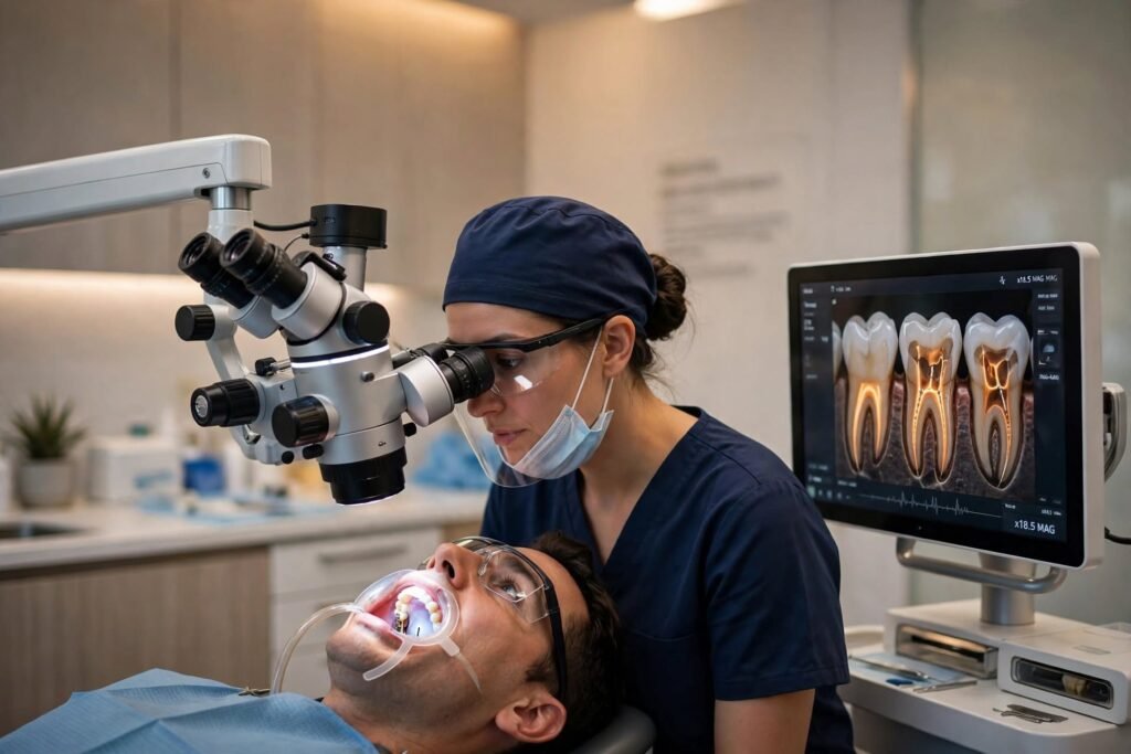
What microscope dentistry actually is
Microscope dentistry means treating a tooth while looking through a dental operating microscope, rather than relying only on the naked eye. The microscope magnifies the tooth and brings a bright, focused light right along the dentist’s line of sight, so the inside of the tooth is lit up instead of sitting in shadow.
Think of it this way. Without magnification, a dentist often has to work partly by feel — following the shape of a canal with fine instruments and checking progress with X-rays. With a microscope, much more of that work becomes visual. The dentist can watch what is happening as it happens, adjust in the moment, and show you images afterwards so the treatment makes sense, not just the final result. That shared view is part of the value: you are not simply told a canal was difficult, you can be shown where it was, why it mattered, and what was done about it during treatment.
Naked eye, loupes and microscope: what is the difference?
Many patients hear about loupes and assume they are the same thing. They are not, although all three approaches have a place in dentistry.
With the naked eye, a dentist can judge the overall tooth, the bite and the restoration well, but fine detail inside a canal is limited. Loupes — small magnifying lenses worn like glasses — are a step up. They help with general precision and posture, and many dentists use them for everyday work.
A dental operating microscope goes further for endodontic work. It gives a stable, magnified and well-lit view deep inside the tooth, which matters most when the problem is hidden: an extra canal tucked in an unusual place, a hairline crack, a canal that has narrowed or calcified, or a tiny broken instrument from earlier treatment. Those details can decide whether a tooth can be treated thoroughly, so the difference is not just comfort — it is visibility where it counts. For a patient, that extra visibility can also mean a clearer explanation of why a tooth needs specialist care, why it may take longer, or why it may not be treatable at all.
What the microscope allows the dentist to do
The clearest benefit is finding what might otherwise be missed. A microscope helps the dentist look for additional canals or canals with an unusual shape, check the floor of the tooth for small cracks, and follow curved or narrow canals more carefully. If a canal was missed in an earlier root canal, that extra visibility is often why a specialist assessment is suggested.
It also helps with how much tooth is removed. When you can see more clearly, you can aim to remove infected or damaged tissue precisely while preserving more healthy tooth structure. That matters, because the strength of the treated tooth afterwards depends partly on how much sound structure remains and how well it is restored.
Finally, the microscope helps the dentist check the work before finishing — whether canals look thoroughly cleaned, whether the filling material has sealed the space as planned, and whether anything still needs attention. In many cases the procedure can also be documented with photos or video, which helps you understand what was found and why a particular step was needed. It also supports careful record-keeping, so if you need follow-up or a second opinion later, there is a clearer picture of what was found, what was treated, and what still needs to be watched at review appointments.
Beyond root canals: where else magnification helps
Root canal treatment is where a microscope is talked about most, and for good reason — that is microendodontics, the specialist area of Dr. Pritesh Agrawal at Phoenix. But careful magnification is useful elsewhere too.
It can support restorative work, where the edge where a filling or crown meets the natural tooth needs to be neat and well sealed. It can help in cosmetic dentistry, where small details affect how a restoration looks and fits. It can also assist with periodontal and implant-related precision, and with spotting subtle problems earlier during an examination.
That does not mean a microscope alone decides the outcome. Materials, technique, the final restoration and your own care at home all still matter — a point we return to below. Used well, magnification is one part of careful, well-planned care, not a substitute for it.
What you will experience as a patient
From your side of the chair, treatment under a microscope will feel familiar in most ways. You will still have a normal examination and X-ray first, local anaesthesia where needed, and the usual protective measures during treatment. What you may notice is a more focused, unhurried pace, especially in a complex case.
Complex microscope work can take longer than a straightforward appointment, because finding and cleaning hidden anatomy takes time. Some patients also find it reassuring to see an image of their own tooth on a screen and have the dentist point out what was found, rather than hearing only a verbal summary.
You may also be told that a microscope case can cost more than a standard case. That depends on the tooth, its complexity, the time and specialist skill involved, and the restoration needed afterwards. Phoenix does not publish a single fixed price for this, so your fee should be explained after examination, before treatment begins.
When a microscope helps most — and what it cannot do alone
A microscope is especially helpful for complex teeth, calcified or curved canals, retreatment of a root canal that has not settled, suspected cracks, and cases where something was missed or left behind earlier. In a straightforward case it still offers a clear view, but its value is greatest when the anatomy is difficult.
It is not the whole answer by itself. A well-cleaned canal still needs a good seal and a sound final restoration, and the tooth still needs daily hygiene and follow-up. If a tooth is cracked in a way that cannot be restored, magnification will help show that clearly — it will not make an unrestorable tooth restorable.
Conclusion: ask whether your tooth needs a closer look
Microscope dentistry is, at its heart, about not guessing inside a tooth that is too small to judge by eye alone. If you have a complex root canal, a tooth that still troubles you after earlier treatment, or you have simply been told your case is difficult, it is worth asking whether microscopic assessment would help.
Phoenix Multispeciality Dental Clinic in Bardoli, Gujarat, is the first and only clinic in Bardoli to provide dental treatment under a microscope. To discuss your tooth, bring any previous X-rays and treatment notes to a consultation at Phoenix in Bardoli, and ask what can be seen, what can be treated, and what the plan is if the tooth cannot be saved.
Frequently asked questions
1. What is microscope dentistry?
Microscope dentistry is dental treatment carried out while the dentist looks through a dental operating microscope. It magnifies the tooth and lights the working area, so fine details inside the tooth — such as extra canals or small cracks — are easier to see and treat carefully.
2. Is microscope root canal treatment better than a normal root canal?
It offers a clearer, magnified view, which can be especially helpful in complex, curved, calcified or retreatment cases. Whether it is the right approach for your tooth depends on examination and X-ray. It does not guarantee a particular result, because the condition and restorability of the tooth also matter.
3. Does microscope dentistry hurt more or less?
The microscope itself does not add pain. Treatment is carried out with local anaesthesia where needed, in the usual way. Any soreness afterwards depends more on the condition of the tooth and the treatment carried out than on the microscope, so discuss comfort and aftercare with your dentist beforehand.
4. How long does a microscope root canal take?
It varies by tooth and complexity. A straightforward case may be completed more quickly, while a tooth with hidden, curved or calcified canals, or a retreatment case, can take longer and may need more than one visit. Your dentist can give a realistic estimate after examination and X-ray.
5. Why might a dentist recommend a microscope for my tooth?
Common reasons include a canal that may have been missed earlier, a narrow or calcified canal, a curved root, a suspected crack, a broken instrument to locate, or a root canal that has not settled. The aim is to see the problem clearly before deciding how to treat it.
6. Can a microscope find a missed canal?
A magnified, well-lit view can help a dentist look for additional or unusually placed canals that are hard to see with the naked eye. Finding a missed canal can be an important part of retreatment, but whether it can be cleaned and sealed still depends on the tooth’s anatomy and condition.
7. Is microscope dentistry only for root canals?
No. Root canals are its best-known use, but magnification can also support restorative work, cosmetic dentistry, periodontal care and implant-related precision, as well as a closer examination of subtle problems. Your dentist will advise where it adds value in your case.
8. Will microscope treatment cost more?
It can, because complex cases take more time and specialist skill, and the final restoration also affects the total fee. There is no single fixed price that fits every tooth. Ask for a clear estimate after examination, including what is and is not included, before treatment starts.
9. Can every tooth be saved with a microscope?
No. A microscope helps the dentist see and treat a tooth more precisely, and to judge restorability more clearly. If a tooth is cracked or damaged in a way that cannot be restored, or has lost too much support, saving it may not be possible, and your dentist should explain the alternatives.
10. Why does Phoenix offer microscope dentistry in Bardoli?
Phoenix Multispeciality Dental Clinic in Bardoli, Gujarat, is described on its website as the first and only clinic in Bardoli to provide dental treatment under a microscope. Its doctor, Dr. Pritesh Agrawal, is a microendodontist, so microscope-guided root canal and precision care are central to the clinic’s work.